Search This Blog

Monday, September 26, 2011

3.18 and 3.19

3.18 Recall the meaning of the terms: dominant, recessive, homozygous, heterozygous, phenotype, genotype and codominance.

and...

3.19 Describe the patterns of monohybrid inheritance using a genetic diagram.


Monday, September 19, 2011

3.24 Mitosis


3.24 Understand that the division of a diploid cell by mitosis produces two cells which contain identical sets of chromosomes.

- Mitosis is a form of cell division which results in growth which occurs by an increase in the number of cells.

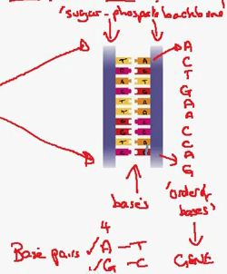
- A normal cell has a nucleus. The number of chromosomes in a nucleus is known as a diploid number which can be abbreviated to the formula 2N. 2N for humans is 46.

- In the process of mitosis this normal cell will divide to form two new cells both with a nucleus. If we take a closer look at the nucleus from each of these cells, we find they both have a diploid nucleus. Meaning they both still have 46 chromosomes meaning they are identical. Sometimes described as daughter cells.

- They are identical because they have the same number of chromosomes and because they have the same set of chromosomes. This means that if we find one chromosome in one of the cells, we will find an identical version of that chromosome in the other cell.

- From this information a number of questions come to mind.
      1) How are the copy of chromosomes made?
      2) How do they separate into the two cells?

These questions are answered in 3.24b


- From 3.24a we have established that a normal cell will copy its chromosomes and divide into two identical cells during mitosis.

- This copy is a process called DNA Replication. In this process each chromosome undergoes a copying process to form an identical copy of itself with all the same genes. These two copies are held together by a central structure called a centromere. These two copies held together by the centromere are called pairs of chromatids.

- This process of DNA Replication takes place in the nucleus while the nucleus is still intact. This means we can't see this process. This is known as the interphase of the cell cycle.

- The process in which the copies of the chromosomes are seperated is dealt with in 3.24c


- Looking at the stages of mitosis:

1) It is during the 'Interphase' of a cell that DNA replication occurs


2) The first sign that a cell is entering mitosis and cell division, is when we see the breakdown of the nuclear membrane. Basically, the nucleus breaks down. This is a phase known as the 'Prophase'. At this point the chromosomes become visible as a pair of chromatids.


3) At this stage the nucleus is gone, and inside the cell a network of protein molecules are present which are known as the spindle, each of which are fibres. They extend from one end (pole) of the cell to the other. During late prophase the chromosome pair will move towards the centre of the spindle and join on to one of the spindle fibres at the centromere.


4) What happens during late phase can be seen in the next stage of mitosis which is known as 'Metaphase.' In the diagram only one pair of chromotids are illustrated. The chromosomes can be seen in the middle attached at the centromere.


5) The next stage is called the 'Anaphase.' Here the fibre (spindle fibre) shortens and pulls one chromotid in one direction towards on of the poles, and one chromatid in the other direction towards the opposite pole. They are moving a part and being separated.


6) The next stage which is the end of mitosis is called 'Telophase.' At thus stage the nucelus begins to reform around the chromosomes at each end of the cell. This will be the new nucleus for the new cell.


7) And finally now the stage called 'Cyto Kinesis' is when the cell splits into two new cells. THIS IS NOT PART OF MITOSIS. The cytoplasm of the original cell splits, and the membrane fuses across the equator to form the new cells. It is important to understand that each of the new cells contain one chromosome.

Sunday, September 18, 2011

3.16 DNA and Genetic information


3.16 Describe a DNA molecule as two strands coiled to form a double helix, the strands being linked by a series of paired bases: adenine (A) with thymine (T), and guanine (G) with cytosine (C).

- The diagram below shows a Gene Loci being expanded to display the double helix structure of the DNA:


- The double helix formation has two strands which seem to be parallel with one another.

- This next diagram shows the DNA molecule in greater detail again:


- The two strands that are opposite one another are known as the "Sugar phosphate backbone". No chemical details for this are required. And in the center between these two sections we find a group of mollecules called bases.

- the 4 different types of bases are known as: adenine (A), thymine (T), guanine (G) and cytosine (C).

- These 4 pair to make: Adenine and Thymine; and then Cytosine and Guanine. A with T and G with C.

A ----- T
G ----- C

- These base pairs join/glue one side of the double helix to the other.

- If you draw your attention to just one side of the double helix you find a pattern of the bases which are in a particular order. This is the order which we call the gene.


- So we have a gene which can be defined as the order of the bases, and the number of the bases. With these two things somehow this codes for the construction of a protein in the cytoplasm. And this is what gives us the characteristic.


3.15 Genes


3.15 Understand that a gene is a section of a molecule of DNA.

- The diagram below shows a molecule of DNA. It shows the characteristic of the Double Helix shape. A section of this molecule is called a gene. This gene carries the information which forms the characteristic of that particular organism. This could be for example blood group, or petal colour in a flower.

- The genes are located in the nucleus and the information is passed to the cytoplasm. Then the genetic information is transformed in the cytoplasm to a protein. It is this protein which controls the production of the characteristic.

- So the information flows from the gene (in the nucleus), to the protein (in the cytoplasm).

3.14 Chromosomes


3.14 Recall that the nucleus of a cell contains chromosomes on which genes are located.

- Chromosomes are the genetic information within a cell. A typical cell would have a nucleus, and when we ipened that nucleus up we would find a number of chromosomes.

- The chromosome is composed of a molecule known as DNA, which forms a shape known as the double helix. Sections of this molecule are called genes, and one chromosome will have thosands of genes.

- Each gene carries the information for the construction of a protein (see 3.16). This protein gives us the characteristic associated with the gene (for example a blood group).

- Different organisms have different numbers of chromosomes. E.g. Cat = 38, Chicken = 78, Chimpanzee = 42, and a Human = 46.

- Chromosomes operate in pairs (homologous pairs). The diagram illustrates an example of some of the homologous pairs. Homologous nature is based on the length of the chromosomes.


This next diagram shows that if we locate a gene on any position on the chromosome. It will give us a position known as a "Gene Loci". If we go to the same position on the homologous pair we will find the same gene. So for instance we will two versions of each gene for one characteristic. These versions are called "Alleles" (covered in 3.17).

3.13 Secondary Sexual Characteristics


Recall the roles of Oestrogen and Testosterone in the development of secondary sexual characteristics.

- This is addressing a process known as puberty which is about the conversion from child to adult. Not to be confused with adolescence (development of the brain).

- In males the hormone producing these changes is Testosterone. The endocrine gland for this hormone is the testis. Some of the effects include; increased number of produced sperm cells, growth and development of the sexual organs (penis and testis), development of body hair (pubic hair, armpit hair, facial hair), body mass will also increase (partly increase in muscle mass), voice breaking (sudden deepening of the voice), development of a sexual drive in behaviour.

-  In females the hormone producing changes is Oestrogen. The endocrine gland for this hormone is the ovary. Some of the effects include; beginning of the menstrual cycle (includes ovulation and menstrual period), development of body mass (rounding of body shape at hips), development of body hair (pubic hair, armpit hair), gradual deepening of voice (but no sudden breaking process), growth and development of sexual organs (larger breasts).

These are the effects in males and females of puberty; going from a child to a sexually mature adult.


3.11 Placenta


3.12 Describe the role of the placenta in the nutrition of the developing embryo.

- The unborn child as an embryo is unable to breathe, digest or excrete. So the child obtains all of these things through the placenta.

- The embryo has an umbilical cord which travels out towards the lining of the uterus and spreads out to form the placenta. The placenta grows into the lining of the uterus, however biologically it is not part of the mother. it grows out of the embryo, not the mother, so the blood vessels (artery's and veins) inside the placenta are the child's.

- Seeing as the placenta grows into the lining of the uterus it is close to the mothers blood stream. The mother will have glucose, amino acids and fats in her blood stream so some of these cross over into the placenta and travel up the umbilical cord and into the child. These things will go from the mothers blood to the child's blood. This is made more efficient by the adaptations of the placenta including a large surface area and thin walls.

- So the nutrients a child obtains during embryo development come from the mother. As well as this the child produces molecules like carbon dioxide and urea which are sent in the opposite direction and excreted through the mother.

3.10 Menstrual Cycle


3.10 Understand the role of oestrogen and progesterone in the menstrual cycle.

- Oestrogen and Progesterone are both hormones. Hormones are produced in a structure known as an endocrine gland in the ovary. Hormones travel through the blood from this gland to its target tissue. This is where the hormone will have it's effect. Sometimes even multiple effects.

- The Ovary (as shown in diagram) is the endocrine gland for oestrogen.

1) The first effect of Oestrogen takes place in the lining of the uterus. Oestrogen is sent through the blood stream to the endometrium.  The diagram shows the difference in the thickness of the lining of the uterus. In the first half of the cycle the lining becomes increasingly thick.

2) The second effect of Oestrogen is the release of a hormone named LH. This is caused when Oestrogen flows through the blood stream to the brain. The peak of this hormone LH is about day 13 in the cycle which causes the ovary to release an egg. This is when Fertilisation is now possible to occur.

Closer look at what happens in the ovary:

- During the first 13 days of the cycle a structure called a follicle begins to grow larger and larger. The center of this follicle is the egg. It is the cells around the follicle which produce the Oestrogen. Around about day 13 the follicle reaches its greatest size at the same time as LH causes the wall of the ovary to rupture. This releases the egg into the oviducts. This process is called Ovulation.

- Once this process is complete the now empty follicle changes function and begins to turn yellow. This gives us the new name Corpus Luteum. This is known to produce Progesterone, so Corpus Luteum is the endocrine gland for Progesterone. This hormone travels through the blood stream to the lining of the uterus. This is effect 3 which makes sure that the lining of the uterus does not break down. It maintains the lining of the uterus. In this condition it is possible for the fertilised egg to implant into the wall and develop into a pregnancy (between the two lines left and right of number 3 on diagram)

- However if this does not occur the Corpus Lutiem breaks down and Progesterone levels fall. This results in effect number 4 which is the break down of the lining of the uterus. This is known as the menstrual period or bleeding. This marks the end of one menstrual cycle and the beginning of another.


3.9 Male Reproductive System

3.9 Recall the structure and function of the male and female reproductive systems.


Male Reproductive System:


- Bladder: Store urine.

- Testis: To carry out the process called meiosis which produces the gamete called sperm cells.
- Epididymis: Store sperm cells.

- Vasdeferens: To carry sperm cells to the penis during sexual stimulation. Tube pulses and pushes the sperm cells along the tube to the penis.

- Prostate: Adds about 20% to 30% of the volume of semen and is known to contain sugars and alkali. The alkaline nature of the prostate secretions neutralise the acidic secretions within the vagina.

- Seminal Vesicles: Also produce sugar based secretions which are also alkaline. Make up around about 70% of the semen in which the sperm cells can travel.

- Urethra: Common tube which joins the left and right testis. Carries semen and urine down to the penis.

- Penis: Carry sperm cells into the vagina during sexual intercourse.


Female Reproduction System:


(Note - At this point before pregnancy the uterus structure is no larger than an orange.)

- a) Ovary: Meiosis occurs to produce eggs (female gametes)

- b) Oviducts: Carries eggs to uterus. Also the location at which Fertilisation takes place.

- c) Uterus: Wall of the uterus is made of muscle. This is the part which will stretch to accommodate a pregnancy, and would contract during birth.

- g) Lining of the Uterus: Accepts and develops the fertilised egg into the embryo here. Also development of the placenta which implants into the lining of the uterus.

- d) Uterus Space: Where the baby develops.

- e) Cervix: Entrance to the uterus. Here is where the sperm cells will make their way up into the oviducts.

- f) Vagina: Where the penis is inserted and sperm cells are sent into the cervix. The vagina collects the sperm cells and allows them to make their way up into the uterus.

3.2 Fertilisation


3.2 Understand that Fertilisation involves the fussion of a male and female gamete to produce a zygote that undergoes cell division and develops into an embryo.

- Process begins with the adult male and female. The cells which make up an adult are described as "diploid" meaning it has a complete set of chromosomes which for humans is 46.

- In the male the cells are divided into sperm cells by meiosis which only has 23 chromosomes. Meiosis halves the number of chromosomes in the cell turning it from a diploid cell to a haploid cell. The same happens in the female cells.

- In sexual reproduction, these two cells are joined/fused together so that they form one cell. This process is known as Fertilisation which involves the combining of two haploid cells (23 chromosomes each) to form a new diploid cell with 46 chromosomes known as a "zygote". This new cell has the same number of chromosomes as any other regular adult, which is a combination of the male and female chromosomes.

- The newly formed Zygote now goes through a process called Mitosis. This is when the new cell divides to form two cells. Both cells will still have 46 chromosomes. These two cells would then divide again in the same way until their are sufficient cells for the structure to be called an embryo.

3.1 Sexual and Asexual reproduction


3.1 Describe the differences between sexual and asexual reproduction.

Sexual Reproduction:
- Sexes exist (can identify male/female)
- Cells called gametes are produced (sperm cell and egg cell)
- Meiosis (type of cell division; halves the number of chromosomes in the gamete cell)
- Fertilisation (sperm cell and egg cell fuse together)
- Large Variation in the population. Sexual reproducing populations have a wide variety or variation (differences).

Asexual Reproduction:
- No sexes
- No gametes
- Mitosis (type of cell division; number of chromosomes is maintained constantly. So a cell with 20 chromosomes will divide to produce two cells each with 20 chromosomes. The two cells are identical)
- No Fertilisation
- Small Variation in the population. Asexual reproducing populations are identical ("clone") with the only variations as a result of mutation.

Monday, September 12, 2011


3.12 Understanding how the developing embryo is protected by amniotic fluid.

- Surrounding the embryo inside the uterus is a fluid which is called the Amniotic fluid.

- One of the main functions of this fluid is protection. It comes from the fact that the fluid cannot be compressed, which means when we try and squeeze the fluid it absorbs the pressure. This means that any blows or force that is applied to the uterus wall will be absorbed by the amniotic wall relieving the pressure and protecting the child.

- A way to experience this would be in a Swimming Pool. If you try to kick your leg in a swimming pool it is difficult to do this at speed and your leg won't move quickly. It is really difficult to generate any force. This is an example of how the amniotic fluid protects the unborn child.

- Another way in which the amniotic fluid protects the child is by keeping it floating in the uterus. The unborn child has no calcified bones to support its weight so it is important that the fluid prevents the child from bearing weight on it's bones.


3.11 Describe the role of the placenta in the nutrition of the developing embryo.

- When the child is in the uterus; which is a water filled environment containing amniotic fluids, the child cannot digest, breath or excrete. To obtain nutrition the embryo has an umbilical cord which grows out of the embryo and leads to the Placenta.

- Note: The placenta grows out of the embryo and not the mother. The blood vessels inside the placenta are the child's blood vessels (arteries and veins)

- Placenta grows into the wall of the uterus.

- The Mother continues to eat during pregnancy which means in her bloodstream there will be the normal Glucose, Amino acids and Fats. This will travel through her blood stream and into the wall of the uterus. From there they will cross into the child's bloodstream and into the child. This crosses at the Placenta. (reason why the Placenta has a large surface area and thin wall).

- Nutrients that the child receives as an embryo comes from the mother. The child also produces molecules which are exchanged back into the mothers blood which includes things like Carbon Dioxide.